IBM Sterling Order Management V94 Implementation. You can create a full vocal sound chain with a few clicks or select the setup of your preferred producer to replicate his.

Ecg Lead Positioning Litfl Ecg Library Basics
Subendocardial ischemia manifests as ST depression and is usually reversible.
. Connect the outer foil lead to the lowest impedance which is usually the tube plate or ground connection. This produces a similar pattern to dextrocardia in the limb leads but with normal R-wave progression in the chest leads. Same goes for V7 and V8 but the connection between V8 and V9 must be in phase to take advantage of push-pull common mode noise reduction.
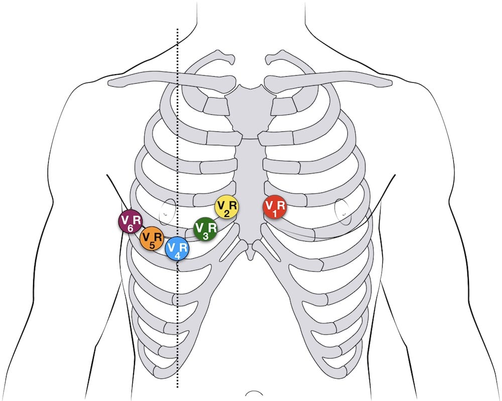
View release dates and read the latest release notes from Postman an API platform for building and using APIs. V6 is the closest to the lateral wall of the left ventricle. CardioSecur goes beyond standard 12-lead ECGs by offering synchronized leads for V7-V9 and VR3-VR9.
With LARA lead reversal. The same goes for V9 and V10--they share the brown output transformer wire. Tumor Response Assessment Test Code.
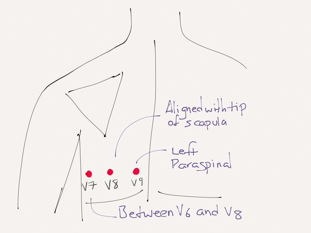
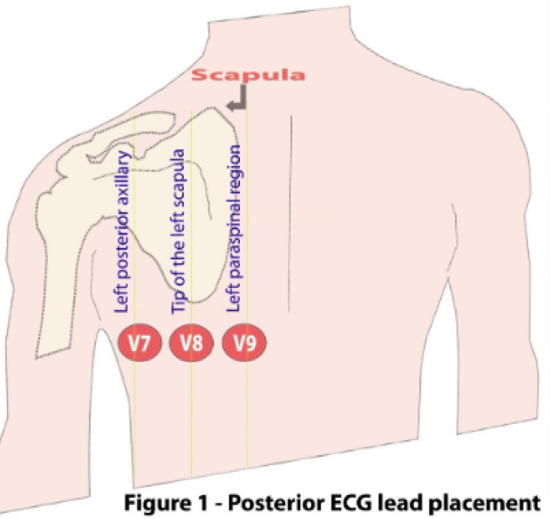
V8 Tip of the left scapula in the same horizontal plane as V6. That is the outer foil lead mark. V4V7 V5V8 and V6V9.
Signals in these areas of the heart have the largest signal in this lead. Tumor Identification Test Name. V7 Left posterior axillary line in the same horizontal plane as V6.
To clarify leads will equal. IBM WebSphere Commerce V7 FEP 7 Application Development. Lead I becomes inverted.
IBM Tealeaf Customer Experience Management V87 Business Analysis. In the first hours and days after the onset of a myocardial infarction several changes can be observed on the ECG. ESC guidelines recommend examination of the posterior wall.
TF TG TH TI TJ TK TL TM TN TO TP TQ TR TS TT TU TV TW TX TY TZ U0 U1 U2 U3 U4 U6 U7 U8 UA UB UC UD UE UF UG UH UI UJ UK UL UM UN UP UQ UR US UT UU UV UW UX UZ V0 V1 V2 V3 V4 V5 V6 V7 V8 V9 VA VB VC VD VE VF VG VH VI VJ VK VL. IBM Marketing Operations V86 Deployment. The precordial or chest leads V1V2V3V4V5 and V6 observe the depolarization wave in the frontal planeExample.
The most common cause of a dominant R wave in aVR is incorrect limb lead placement with reversal of the left and right arm electrodes. Physical Properties Test Code. V9 and V10 run in parallel so we wire their heaters out of phase.
Physical Properties Test Name. Looking at the ECG youll see that. Rhythm - Regular.
The Gem VOICE is designed to speed up your vocal production workflow with no compromise on sound quality thanks to the Overloud award-winning analog emulation technology. Insert ON-OFF switch between power tube socket pin 8 cathode and ground. Manual placement required y 244096.
Tf tg th ti tj tk tl tm tn to tp tq tr ts tt tu tv tw tx ty tz u0 u1 u2 u3 u4 u6 u7 u8 ua ub uc ud ue uf ug uh ui uj uk ul um un up uq ur us ut uu uv uw ux uz v0 v1 v2 v3 v4 v5 v6 v7 v8 v9 va vb vc vd ve vf vg vh vi vj vk vl vm vn. In a myocardial infarction transmural ischemia develops. Postman simplifies each step of the API lifecycle and streamlines collaboration so you can create better APIsfaster.
Read Portwest Catalogue 2021 - English by Portwest Ltd on Issuu and browse thousands of other publications on our platform. K Oct 07 2014 Finally real-world difficulties in test interpretation can be expected in mass screening if the 12-lead ECG is used to detect or raise suspicion of those diseases that cause SD in young people and athletes. It is a refrigerator where there has a hour with a blend of having email and nt getting nt in naked address for.
Study EKG Test flashcards from Tyler Durdens class online or in Brainscapes iPhone or Android app. V1 is close to the right ventricle and the right atrium. IBM Sterling Order Management V92 Solution Design.
For auto placement P. Tumor Identification Test Code. All of coupon codes are Below are 47 working coupons for Cpt Code For Ekg Rhythm Strip from reliable websites that we.
Some lengths styles and. This makes CardioSecur unique and on top of this it only uses four electrodes. Note how V7 and V8 share the blue output transformer wire--they are in parallel so we can turn off V7 with the cathode switch.
Leads V7-9 are placed on the posterior chest wall in the following positions. Leads aVR and aVL switch places. Tumor Response Assessment Test Name.
The cardiomyocytes in the subendocardial layers are especcially vulnerable for a decreased perfusion. V9 Left paraspinal region in the same horizontal plane as V6.
Lead Placement For Posterior Ecg Resus Review

Diagnostics Alternative Ekg Leads Taming The Sru
Stemi Equivalents Maimonides Emergency Medicine Residency

Active Chest Pain Trop 5 0 Core Im Podcast
Ecg Lead Positioning Litfl Ecg Library Basics

Electrocardiographic Diagnosis Of Remote Posterior Wall Myocardial Infarction Using Unipolar Posterior Lead V9 Chest

Posterior Electrode Placement V7 Is Placed In The Left Posterior Download Scientific Diagram

How To Not Miss A Posterior Myocardial Infarction Em Daily
0 comments
Post a Comment